Animal Tissues Identification
Explore the fascinating world of animal tissues. Learn to identify and differentiate between various types of tissues based on their structure, function, and location in the body.
Help & Instructions
▼- Tissue Identification: Identify tissue types based on microscopic images
- Tissue Characteristics: Match tissues with their functions and locations
- Use the hint button if you need help with identification
- Try different difficulty levels to challenge yourself
- Identify four main types of animal tissues
- Understand the structure and function of each tissue type
- Recognize tissue locations in the body
- Differentiate between epithelial, connective, muscular, and nervous tissues
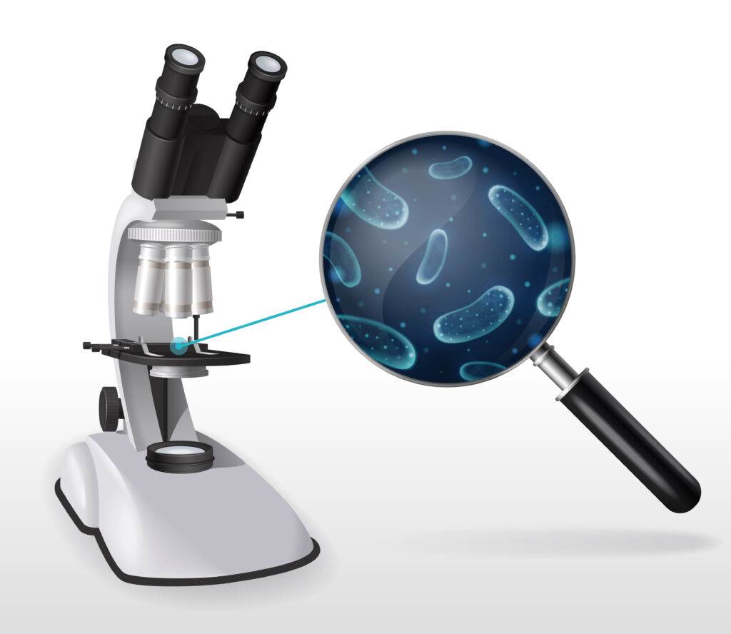
Tissue Identification: Microscopic View
Identify the type of animal tissue shown in the diagram.
Tissue Characteristics: Function and Location
Match each tissue type with its correct function and location in the body.
Animal tissues are groups of cells with similar structure and function that work together to perform specific tasks. The four main types are epithelial, connective, muscular, and nervous tissues. Each has unique characteristics that make it suitable for its specific role in the body.
The Science of Animal Tissues
Four main types of animal tissues:
- Epithelial Tissue: Covers body surfaces, lines cavities, and forms glands
- Connective Tissue: Supports, binds, and protects organs (bone, blood, cartilage)
- Muscular Tissue: Responsible for movement (skeletal, cardiac, smooth)
- Nervous Tissue: Transmits nerve impulses (brain, spinal cord, nerves)
Each tissue type has distinctive characteristics:
Closely packed cells, basement membrane
Cells scattered in extracellular matrix
Elongated cells with striations
Neurons with dendrites and axons
Understanding where tissues are found and what they do:
- Epithelial: Skin (protection), lining of digestive tract (absorption)
- Connective: Bones (support), blood (transport), fat (storage)
- Muscular: Skeletal muscles (voluntary movement), heart (pumping blood)
- Nervous: Brain (coordination), nerves (signal transmission)
Learning to identify tissues helps students understand how the body is organized at the microscopic level. This knowledge forms the foundation for understanding organ systems, physiology, and how the body maintains homeostasis.